News & Blogs 2023-12-06
Introduction and selection of secondary antibodies

EN
CN
EN
News & Blogs 2023-12-06
Introduction and selection of secondary antibodies
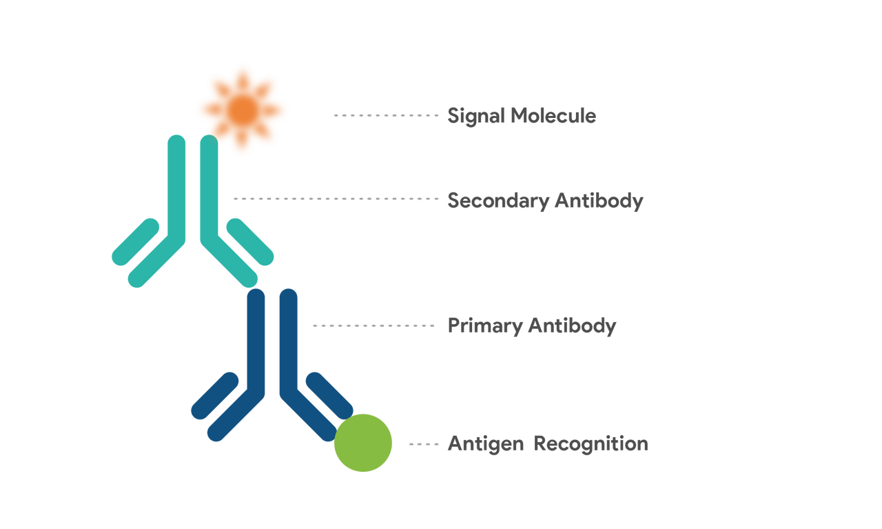
A secondary antibody serves to bind specifically to a primary antibody. It is frequently employed in various immunological assays like WB, ELISA, and immunofluorescence to identify target proteins and magnify the primary antibody's signal.
Secondary antibodies capitalize on antibodies' large protein structure with antigenic traits to inoculate a different species, stimulating its immune system to generate immunoglobulins directed against the primary antibody. These secondary antibodies react against all antibodies of a specific species (e.g., IgG, IgM, or IgA) (e.g., mice), amplifying their utility in diverse experiments.

A primary antibody is designed to target an antigen, while a secondary antibody specifically targets a primary antibody. In essence, antibodies can play a dual role as both antibodies and antigens, triggering the body to create antibodies in response. When an antigen enters the body, it prompts the immune system to generate an immune response, allowing B cells to produce unique proteins that bind precisely to the related antigen. Both primary and secondary antibodies possess the ability to bind to other substances, with a primary antibody capable of binding to at least two other groups (the substrate and the secondary antibody).
Primary antibodies are adept at binding specifically to the substrate antigen, making them capable of recognizing the target. However, their interaction with the substrate isn't visible to the naked eye. In experiments, primary antibodies containing their own visible markers can be used independently without the need for secondary antibodies. Nevertheless, this approach can be expensive and labor-intensive due to the primary antibody's recognition limited to one substrate.
Many secondary antibodies are equipped with markers like fluorescent, radioactive, chemiluminescent, or chromogenic groups, enhancing their detectability. These secondary antibodies bind to the primary antibody, assisting in its detection. They offer numerous advantages: regardless of the primary antibody used, employing a secondary antibody can facilitate the replacement of various markers. Moreover, secondary antibodies bind more labeled dyes at the antigen site compared to directly labeled primary antibodies, amplifying the detection signal. This strategy avoids the complexity and cost of chemically labeling (coupling) the primary antibody, which might interfere with antigen recognition.

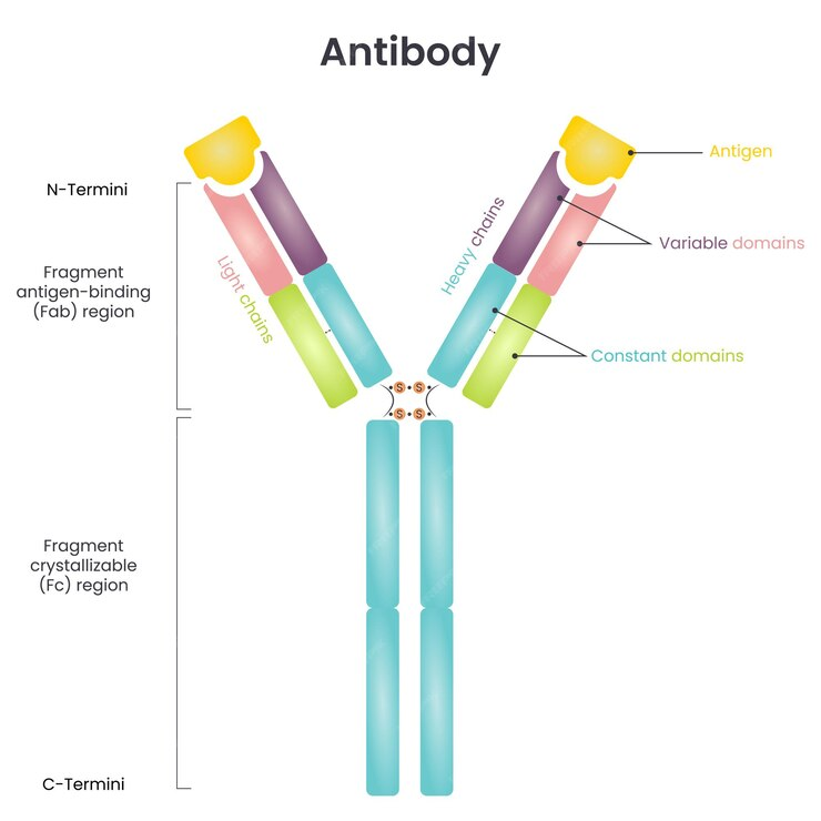
Source: freepik.com
The Fc segment found in antibodies of the same subtype and source remains consistent. Following this principle, secondary antibodies are designed, correlating with the Fc segment subtype of the primary antibody's source.
Matching the isotype of the primary antibody, the secondary antibody is crucially paired. Polyclonal primary antibodies, commonly from rabbit, goat, sheep, or donkey sources, are typically IgG isotypic. Therefore, the corresponding secondary antibody is often an anti-IgG H&L (heavy and light chain) antibody.
1. Selection against the host source of the primary antibody: the secondary antibody points to the species of the primary antibody
2. Selection against the class subtype of the primary antibody: the secondary antibody needs to match the class or subclass of the primary antibody. This is usually for monoclonal antibodies. Polyclonal antibodies are mainly IgG class immunoglobulins, so the corresponding secondary antibody is an anti-IgG antibody.
3.Genus source of the secondary antibody: except for double immunofluorescence labelling method, generally speaking, there is no inevitable connection between different genus sources and the quality of the secondary antibody, and there is not much difference between the secondary antibody from goat and donkey in general experiments.
4. Coupling labels of secondary antibody: Different labels are selected for different experimental methods. Generally speaking, the probes coupled to secondary antibody mainly include enzymes (horseradish peroxidase HRP and alkaline phosphatase AP or its derivatives APAAP, PAP), fluorescent groups (FITC, Rhodamine, Texas Red, PE, Rhodamine, Dylight, etc.), biotin, gold particles. Biotin, gold particles. Generally, HRP-labelled secondary antibodies are recommended for WB, ELISA, IHC, etc., and FITC-labelled antibodies are recommended for IF FC.
5. Selection of pre-adsorbed secondary antibody: For immunoglobulin-rich tissues and cells, it is usually recommended to use pre-adsorbed serum secondary antibody for protein blotting. Pre-adsorbed secondary antibodies have a lower probability of interacting with endogenous immunoglobulins, thus reducing the non-specific background.
6. Special requirements: The F(ab')2 fragment form of the secondary antibody avoids binding to Protein A/G or to the Fc receptor, and the molecule itself is relatively small, making it easier to penetrate into cellular tissues with higher sensitivity. The Fab fragment form of the secondary antibody contains only one binding site, which can be used to sequester endogenous immunoglobulins; generally, no requirement is required. Anti-IgG H&L, which is the most commonly used and cost-effective form of secondary antibody, reacts with both the heavy and light chains of the IgG molecule, as well as other immunoglobulin families (IgM, IgA, IgD, IgE) and subfamilies, since all immunoglobulins have the same light chain.
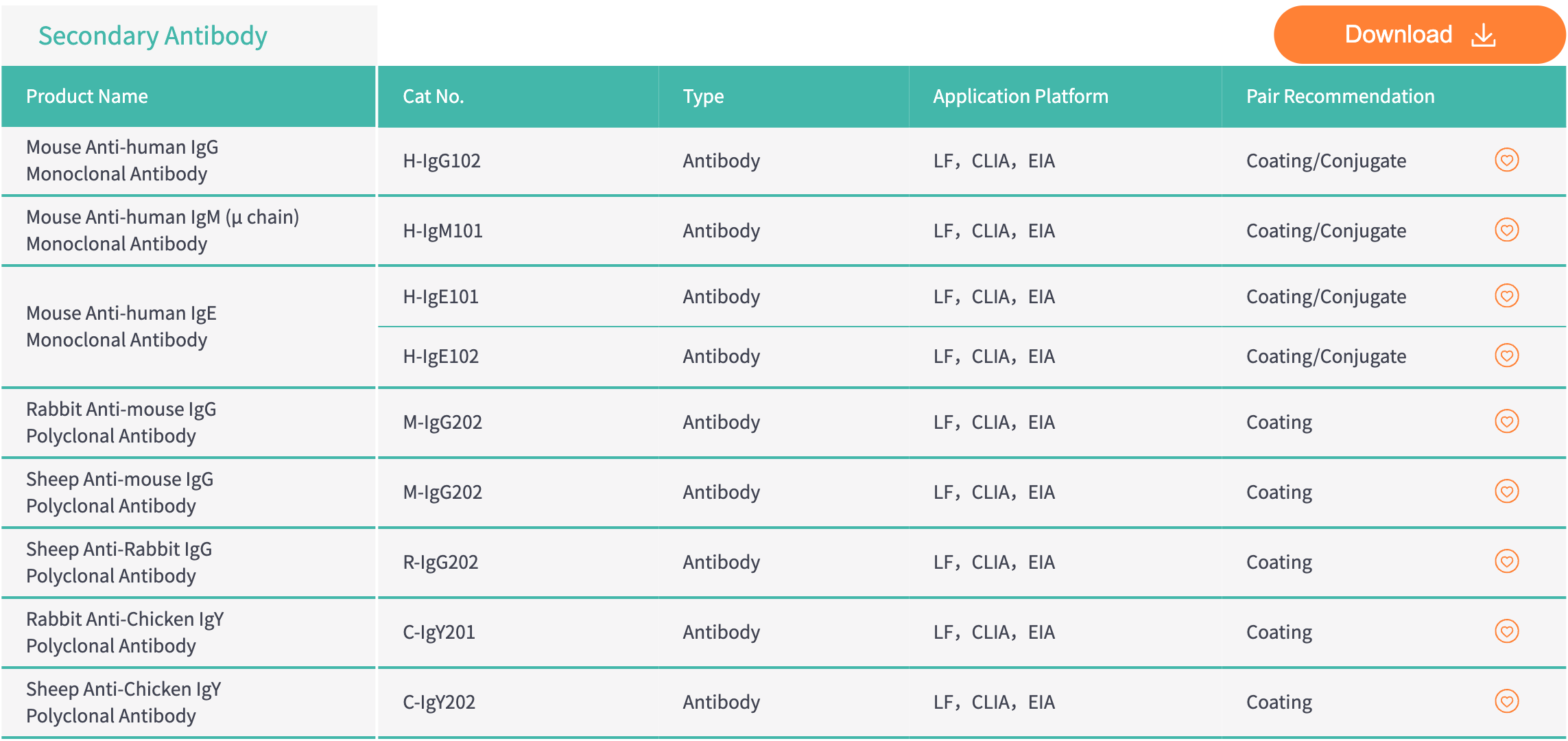
BIOEAST's Solution | Secondary Antibody used in LF, CLIA, EIA
